
The Versatile X-ray Probe: Full-Field Imaging in X-ray Microscopy
X-ray microscopy, characterized by its three-dimensional, non-destructive, and perspective imaging capabilities, is widely applied in fields such as material structure characterization, semiconductor manufacturing defect detection, and aerospace applications.
Click the blue text to follow us
1
Introduction
Top-Unistar
Since Wilhelm Conrad Röntgen discovered X-rays in 1895, over a century of development has brought significant advances in the generation, modulation, detection, and corresponding analytical methods and algorithms of X-rays. The versatile X-ray plays a pivotal role in astronomy, high-energy astrophysics, material analysis in scientific research, quality control and flaw detection in industrial production, security inspection, and medical applications. The analytical methods involved include X-ray diffraction, fluorescence, scattering, absorption imaging, phase-contrast imaging, photoelectron spectroscopy, and ptychography (PTY) imaging. As the most advanced X-ray sources, synchrotron radiation facilities and free-electron lasers (FELs) provide extremely high brightness, supporting cutting-edge scientific research and characterization needs in high-end manufacturing, and creating conditions for the development of new characterization methods.
However, synchrotron radiation sources are costly and have limited beam time resources, making them difficult to widely adopt. Therefore, the scientific research and industrial communities have developed various specialized laboratory instruments based on their respective needs, such as medical CT, industrial and research-grade micro/nano CT, and laboratory X-ray absorption fine structure (XAFS) spectrometers. This development trajectory demonstrates a technological evolution trend from synchrotron radiation to laboratory instruments.
X-ray microscopy, due to itsthree-dimensional, non-destructive, and perspective imagingcharacteristics, is widely applied in fields such as material structure characterization, semiconductor manufacturing defect detection, and aerospace applications. In previous articles"The Versatile X-ray Probe: X-ray Microscopy"and"The Versatile X-ray Probe: Defect-Assisted Imaging in X-ray Microscopy", we introduced themain technical routes of X-ray microscopy: projection geometric magnification, scanning transmission microscopy based on Fresnel zone plates, full-field transmission microscopy, and CDI/PTY techniques. We also detailed the research progress of laboratory X-ray microscopy based on spatial geometric magnification and the intriguing defect-assisted imaging technique.
Today, we will introduce the laboratory full-field transmission X-ray microscopy technique based on spatial geometric magnification and its latest advances.
2
Full-Field Transmission X-ray Microscopy:
From Synchrotron Radiation to the Laboratory
Top-Unistar
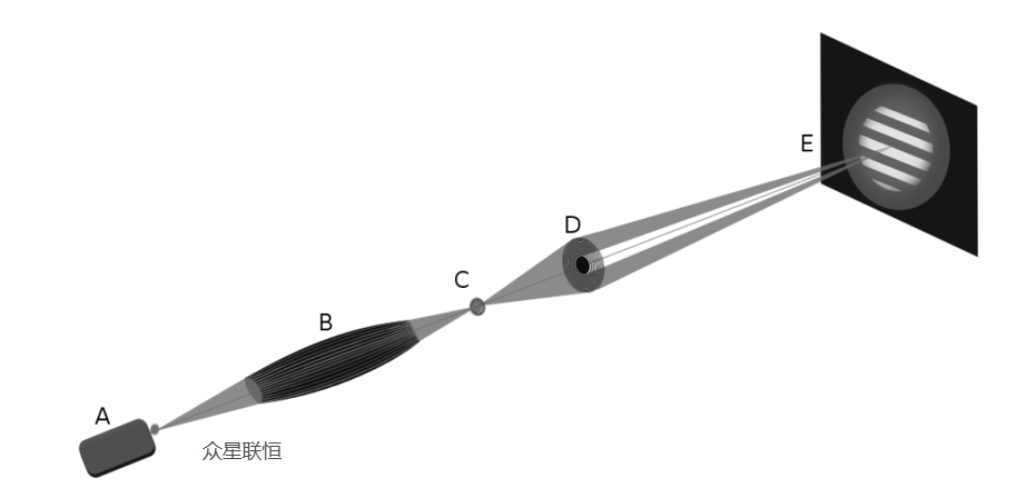
Figure 1 Optical path of full-field transmission X-ray microscopy. Light source (A); polycapillary lens (B); sample with pinhole (C); zone plate (D); detector (E)[1]
As shown in Figure 1 above, in a laboratory full-field transmission X-ray microscopy system, we typically use a high-brightness laboratory light source (liquid metal jet or rotating anode source) to replace the expensive and less accessible synchrotron radiation source. Single or polycapillary optics can serve as X-ray condensers, effectively collecting X-rays from the point source and focusing them onto the sample plane, as illustrated in Figure 1 above. Combined with a high-efficiency Fresnel zone plate and a high-resolution camera at the downstream end, sub-micrometer spatial resolution can be achieved under laboratory conditions.[1]。
Here, we must mention a key figure who brought full-field transmission X-ray microscopy into the laboratory—Dr. Wenbing Yun. As the founder of Sigray (now owned by Applied Materials), a laboratory X-ray system supplier, and Xradia (now owned by Zeiss), an X-ray microscope company, he grew up in a small farming and herding village in Inner Mongolia in the 1960s and 1970s. In 1986, Dr. Yun completed his studies in the Physics Department at Stony Brook University, where he earned his PhD in coherent X-ray diffraction imaging. After graduation, he first joined Argonne National Laboratory and then moved to the Advanced Light Source at Lawrence Berkeley National Laboratory. In 2000, Dr. Yun founded Xradia, dedicated to developing laboratory X-ray microscopes for semiconductor manufacturers. In the summer of 2013, Carl Zeiss acquired Xradia. In 2026, Applied Materials of the United States acquired Sigray.[2]。
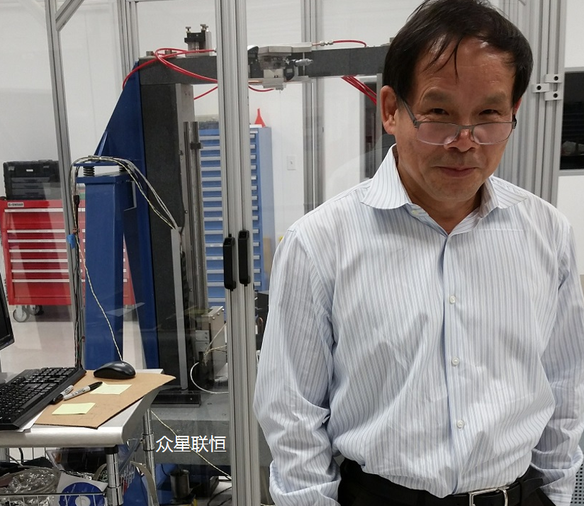
Figure 2 Wenbing Yun: Pioneer of X-ray imaging, founder of Sigray and Xradia. (Sigray)
3
Core Component-Driven Development:
Advances in Full-Field Transmission X-ray Microscopy
Top-Unistar
The core advantage of the polycapillary X-ray lensas a condenser lies in its unique microchannel array structure composed of hundreds of thousands of individual capillaries, which enables Köhler-like uniform illumination, avoiding projection of the source structure onto the sample; supports multi-angle illumination, allowing easy switching between bright-field and dark-field imaging modes by adjusting the beam stop; and has low requirements for spatial coherence of the source, making it easy to pair with conventional laboratory X-ray sources. Its main disadvantages are: the numerical aperture of the polycapillary and the downstream zone plate is difficult to match perfectly, causing some light to be lost and not participate in imaging; it cannot follow the lens equation to compress the source energy to the extreme, so the peak radiation density at the sample plane is lower than that of a precisely designedsingle-capillary system; and the destruction of beam coherence caused by multiple reflections limits its support for advanced quantitative phase imaging.[3-4]。
As shown in Figure 1 above, in 2015, a research team from the University of Applied Sciences Koblenz, Technical University of Berlin, and Max Born Institute in Germany successfully built a laboratory 8 keV full-field transmission X-ray microscope with bright-field and dark-field imaging capabilities, using a 30 W low-power copper target microfocus X-ray source (50 μm), a polycapillary X-ray lens as the condenser, a Fresnel zone plate as the imaging objective, and a direct-detection CCD camera (pixel size 20 μm × 20 μm) as the imaging detector.Figure 3 below shows the system resolution measurement results. By testing the X-ray bright-field image of a 128-sector Siemens star (260× magnification, 77 nm/pixel, 380-minute exposure) and measuring the distance over which the intensity in a specific region changes from 10% to 90%, the results showed an edge spread of 210 nm, indicating a system spatial resolution of 210 nm. Although the theoretical resolution of the zone plate used was 61 nm, the measured 210 nm indicates that the current resolution is mainly limited by the detector pixel size and system matching, rather than the objective lens itself [2]. Regarding system improvements, the authors mentioned that if amicrofocus rotating anode high-brightness X-ray sourcewere used, the exposure time for dark-field imaging is expected to be reduced to 10 minutes or even shorter. Combined with a high-brightness source and a lens-coupled X-ray camera with higher spatial resolution, the resolution could be improved to around 61 nm.[3]。
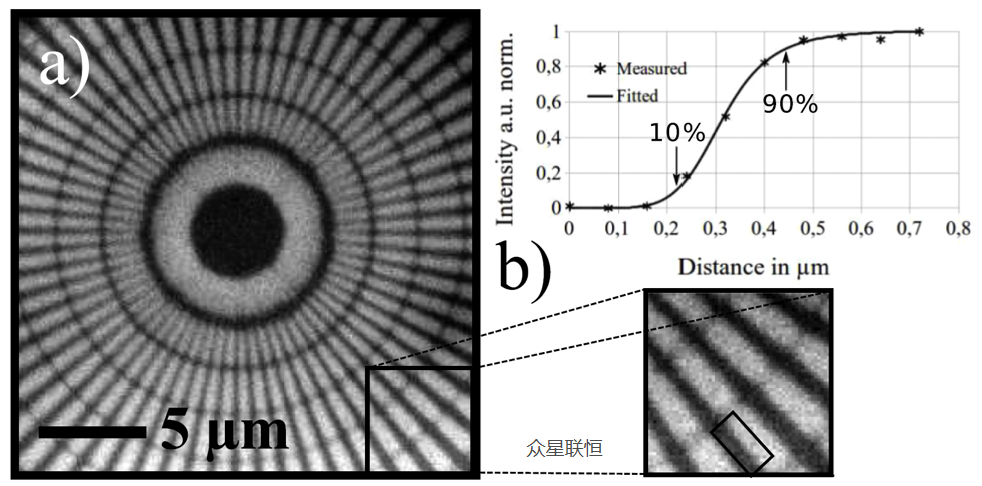
Figure 3. Bright-field image of a Siemens star taken at 260× magnification with an exposure time of 380 minutes (a); imaging resolution evaluated by edge sharpness, with a spatial resolution of approximately 210 nm (b).
As shown in Figure 4 below, in 2016, a research team from Julius-Maximilians-Universität Würzburg and the Fraunhofer Development Center X-ray Technology in Germany used a liquid metal jet high-brightness X-ray source, a polycapillary X-ray lens manufactured by XOS (a partner of Top Unistar) in the United States as the condenser, and a zone plate with a lens-coupled high-resolution CMOS camera to build a 9.25 keV full-field X-ray microscope.
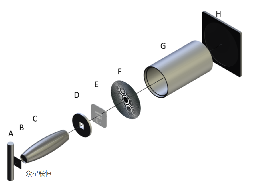
Figure 4 Schematic of individual components of the laboratory-developed X-ray microscope (from left to right): A) liquid metal X-ray source, B) zinc foil, C) condenser, D) pinhole, E) sample, F) Fresnel zone plate, G) vacuum tube, H) detector
To test the system performance, the researchers testedgratings with a 2.4 μm period and 15 μm gold height manufactured by MicroWorks (a partner of Top Unistar) in Germanyusing LIGA technology, as well as 1–3 μm line pairs self-fabricated on 1.5 μm gold foil using focused ion beam. By fitting the grating edge, the edge spread function and MTF curve were obtained as shown in Figure 5 below. At 10% contrast, the system achieved aresolution of 1400 lp/mm (corresponding to a spatial wavelength of 714 nm)。
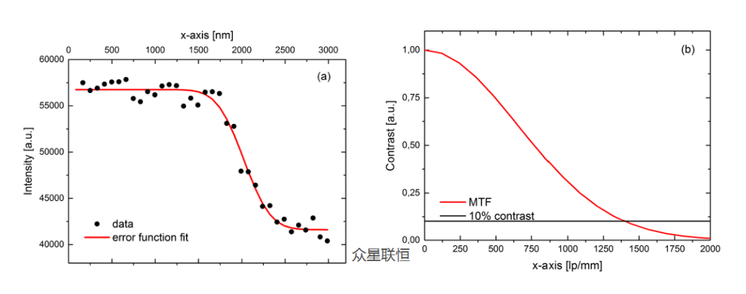
Figure 5 Curve along the grating edge (a). The red line is the error function fitting curve. Corresponding modulation transfer function (MTF) curve (b): at 10% contrast, the resolution reaches 1400 lp/mm (corresponding to a spatial wavelength of 714 nm).
At the same time, the authors mentioned that the focus of future work is tomodify and optimize the condenser into a single-capillary structureto further increase photon flux and preserve more imaging brightness. After achieving better illumination control,Zernike phase-contrast imaging[5]。
can be introduced by adding a phase plate.As is well known, when building a full-field transmission X-ray microscope (TXM) on an X-ray tube source, on the one hand, image blurring occurs due to Fresnel chromatic aberration; on the other hand, the inherently low photon flux of laboratory sources results in long exposure times. Therefore, to maintain good imaging quality, previous setups typically added filters such as Zn after the source to obtain quasi-monochromatic light. Recently, a research team from the Paul Scherrer Institute in Switzerland and the University of Science and Technology of China introduced an achromatic Fresnel zone plate (New breakthrough! Custom combination of compound refractive lenses and Fresnel zone plates achieves achromatic focusing of X-rays over a wide energy range【4】。
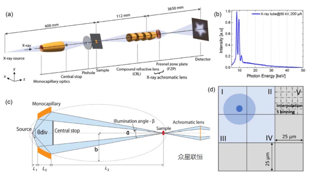
), which had been validated at a synchrotron radiation facility, into a laboratory system, aiming to increase analysis speed and expand the analytical dimensions while maintaining resolution.
Figure 6 Schematic of the laboratory achromatic full-field transmission X-ray microscope. Distances between key components are: source-to-sample distance of 400 mm, sample-to-achromatic lens distance of 112 mm, and lens-to-detector distance of 3650 mm. (b) Measured X-ray energy spectrum of the tungsten microfocus X-ray tube at a tube voltage of 50 kV and tube current of 200 μA. (c) Schematic of the single-capillary optical system and achromatic lens structure (parameters: a=200 mm, b=0.42 mm, β=1 mrad, divergence angle θdiv=13.46 mrad, L1=9.5 mm, LC=10 mm, L2=280.5 mm). (d) Schematic of the MÖNCH pixel detector principle, demonstrating charge-sharing-based interpolation positioning: incident X-ray photons are absorbed within pixel I; the charge cloud diffuses to pixels I–IV, forming a 2×2 pixel cluster and being collected; subsequently, the incident photon position is determined to a virtual sub-pixel level via an interpolation algorithm.As shown in Figure 6 above, the researchers built the first experimental achromatic full-field microscopy system using a specially fabricated single-capillary X-ray lens, an achromatic X-ray lens, and a MÖNCH hybrid silicon pixel detector (pixel pitch 25 μm). With the current optical configuration, this full-field transmission X-ray microscopy (TXM) system achieves a spatial resolution ofat approximately 10 keV X-ray energy480 nm. At the same time, the authors mentioned that the focus of future work is tofurther improve the fabrication precision of the single-capillary optics and the achromatic lensto enhance the light utilization efficiency and reduce aberrations of the full-field transmission X-ray microscopy (TXM) system. In addition, increasing the frame rate of the MÖNCH detector and shortening the imaging acquisition time are also critical, which can be achieved by upgrading the data acquisition server. Furthermore, building atemperature-stabilized protective enclosureis expected to further suppress vibration and thermal drift effects during long exposures.
With convenient and accessible experimental conditions, this system achieves theintegration of structural characterization and elemental characterization (via the detector's energy resolution capability), providing new research approaches and development opportunities for studies in materials science, environmental science, and biology.[6]。
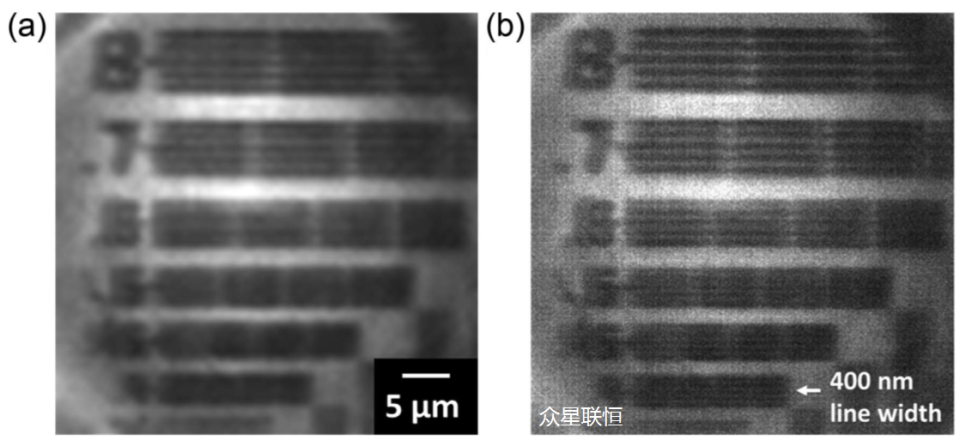
Figure 7 Raw image of the XRnanotech test pattern (a partner of Top Unistar) taken with the achromatic lens. The test pattern contains six groups of line structures with line widths ranging from 400 nm to 800 nm. (b) Image after 5 μm block interpolation, i.e., the original pixels are further divided into 5×5 sub-pixels.
4
Summary
Top-Unistar
In summary, we see that core components such as single and polycapillary X-ray lenses, X-ray Fresnel zone plates, and detectors play a key role in laboratory full-field X-ray microscopy instruments. Higher diffraction efficiency and wider achromatic bandwidth mean making full use of every photon to increase analysis speed; higher-resolution detectors enable higher resolution to reveal finer sample details; pixelated X-ray detectors with high energy resolution add an analytical dimension, bridging structural and elemental characterization. Combined with the story of Dr. Wenbing Yun, we can see that bringing synchrotron X-ray analysis techniques into the laboratory requires not only in-depth understanding or breakthroughs in individual components and the formation of patent barriers, but also a systems engineering approach that requires trade-offs across multiple directions. It also requires consideration of market conditions, team building, and healthy company operations. Undoubtedly, Dr. Wenbing Yun and our partner Torsten Feigl of optiXfab (The hidden giant in Jena, Germany: optiX fab.) are excellent role models who combine scientific research, engineering technology development, and commercialization to the extreme. As domestic scientific instruments flourish today, perhaps we should learn from them.

TOP-UNISTAR
About Top Unistar
As a professional provider of extreme ultraviolet (EUV) and X-ray core components and solutions, Top Unistar offers various core components for full-field X-ray microscopy, such as high-brightness X-ray sources, condensers, Fresnel zone plates, X-ray CCD cameras,high-resolution lens-coupled cameras, and photon-counting, pixelated X-ray detectors. We currently maintain a certain inventory ofEUV and X-ray related equipment and core components, enabling rapid delivery to better serve our users. We have also established a well-equippedX-ray open laboratory, equipped with multiple advanced X-ray source systems, optical components, and detectors, supporting diverse testing, pre-research, and validation work including X-ray imaging, phase-contrast imaging, diffraction analysis, fluorescence detection, and X-ray source performance characterization.
We are a team passionate about EUV and X-ray technology. If you have any questions you would like to discuss with us, please do not hesitate to contact us directly.
References and Materials
Top-Unistar
Baumbach S, Kanngießer B, Malzer W, et al. Setup of an 8 keV laboratory transmission x-ray microscope. Journal of Physics: Conference Series. 2014, 499(1): 012005.
https://analyticalscience.wiley.com/content/news-do/wenbing-yun-life-inspired#media-2
Baumbach S, Kanngießer B, Malzer W, et al. A laboratory 8 keV transmission full-field x-ray microscope with a polycapillary as condenser for bright and dark field imaging. Review of Scientific Instruments, 2015, 86(8).
Korecki P, Sowa K M, Jany B R, et al. Defect-assisted hard-x-ray microscopy with capillary optics. Physical Review Letters, 2016, 116(23): 233902.
https://pubs.aip.org/aip/acp/article/1696/1/020025/814106/Laboratory-source-based-full-field-x-ray
Qu, Di, et al. "Achromatic laboratory full-field transmission X-ray microscope." Photonics Research 14.2 (2026): 392-400.



Content: Kevin & α·Lee
Review: Kevin
Layout: Sylvia
Disclaimer:
Part of the content (including images) in this article is sourced from the internet. The copyright and views of the quoted parts belong to the original authors. Beijing Top Unistar Technology Co., Ltd. publishes and reposts this content to convey more industry information and facilitate online sharing. If you believe this article infringes on your rights, please contact us, and we will address it promptly. If you have any questions, please feel free to contact us at any time.
Welcome to repost; please indicate the source.
FROM READING TO APPLICATION
Put this knowledge to work
Need to assess product suitability, discuss experimental conditions or find technical support? Explore our products and services, or discuss this resource with us.