
Multicapillary Lenses Enable Laboratory-Scale Micro-XEOL Research
Using this background framework, this article focuses on two applications more relevant to the construction of laboratory XEOL platforms: one is the luminescence mechanism and performance optimization of nanoscintillator materials, and the other is XEOL imaging applications for deep biological tissues.
Click the blue text to follow us
In the fields of biology and materials science, X-ray excited optical luminescence (XEOL) is a highly attractive spectroscopy and imaging technique. It uses X-rays to excite scintillating or luminescent materials and monitors their luminescence response in bands such as UV-vis-NIR, thereby revealing the local electronic band structure, defect levels, and energy transfer processes of materials; in biomedical scenarios, XEOL can also leverage the deep penetration capability of X-rays to achieve deeper and lower-background functional imaging than traditional optical excitation. The review by Fan et al. systematically outlines the development of X-ray-excited theranostic technologies from the perspective of nanomaterials, covering directions such as CT contrast-enhanced imaging, XEOL imaging, and multimodal synergistic therapy [1]Using this background framework, this article focuses on two applications more relevant to the construction of laboratory XEOL platforms: one is the luminescence mechanism and performance optimization of nanoscintillator materials, and the other is XEOL imaging applications for deep biological tissues.
(1) XEOL Material Luminescence Mechanism and Performance Optimization
XEOL material research must first answer the question of how X-ray energy is absorbed, transferred, and converted into light by materials. The review mentions that the luminescence performance of scintillating nanomaterials is generally jointly influenced by host matrix composition, crystal phase, doping concentration, and surface properties: host matrices containing high-atomic-number elements are conducive to enhancing X-ray absorption, appropriate activator ion doping can increase radioluminescence intensity, while crystal phase differences, surface defects, and core-shell structures can also significantly alter energy transfer efficiency. Taking systems such as NaGdF4:Eu as examples, the X-ray excited luminescence intensity is closely related to the Eu3+ doping ratio and crystal phase; passivating surface defects with an inert shell layer also helps to increase luminescence intensity. In addition, persistent luminescence nanomaterials can continue to release near-infrared afterglow after X-ray irradiation ceases, and rare-earth-doped nanoprobes can convert X-ray energy into short-wave infrared signals. Therefore, research on the material side is not merely a preliminary step for imaging applications; it itself requires stable, high-throughput, and focusable X-ray excitation conditions for spectral acquisition, material screening, and micro-region luminescence distribution characterization.
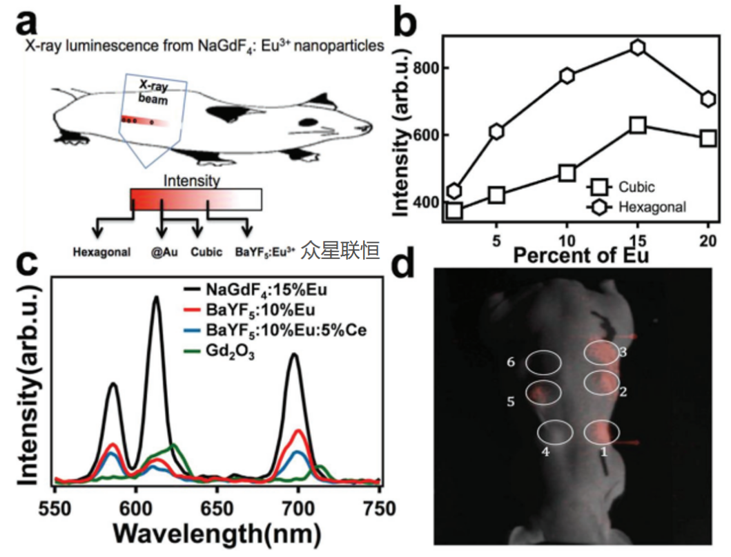
Figure 1. XEOL spectra and imaging validation of NaGdF4:Eu nanoparticles.
a) Schematic of NaGdF4:Eu nanoparticles used for XEOL imaging; b) Comparison of X-ray luminescence intensity of NaGdF4:Eu nanoparticles at different Eu3+ concentrations; c) Comparison of X-ray excited luminescence intensity of nanoparticle host matrices with different Eu doping;
d) X-ray luminescence imaging results: 1) hexagonal-phase NaGdF4:15%Eu; 2) NaGdF4:15%Eu@Au; 3) hexagonal-phase NaGdF4:15%Eu; 4) BaYF5:10%Eu/5%Ce; 5) BaYF5:10%Eu; 6) saline control group without particles.
(2) XEOL Deep Biological Imaging
The core advantage of XEOL imaging lies in breaking through the depth limitation of traditional fluorescence imaging (Seeing the subtle moments of life: high-sensitivity CCD cameras, the ideal choice for in vivo imaging) and completely eliminating background noise. Compared with direct excitation from outside the body using UV-vis-NIR light, X-rays undergo less scattering and attenuation in tissue and can excite scintillating nanomaterials at greater depths; subsequently, the visible, near-infrared, or short-wave infrared light emitted by the materials is collected by the detection system, thereby reducing the autofluorescence and shallow penetration limitations of traditional optical imaging. The review by Fan et al. demonstrates several representative pathways: scintillating nanomaterials such as NaGdF4:Eu can be used for XEOL imaging validation; rare-earth-doped core-shell nanoprobes can generate short-wave infrared signals at approximately 1525 nm under X-ray excitation for lymphatic drainage and sentinel lymph node localization; and persistent luminescence nanomaterials such as LGO:Cr can continue to emit near-infrared afterglow after X-rays are turned off, enabling low-background imaging under tissue coverage. Together, these works illustrate that XEOL imaging is concerned not only with "seeing," but also with how to exploit the emission band, afterglow, and energy transfer characteristics of luminescent materials to obtain higher signal-to-noise ratios and deeper functional information.
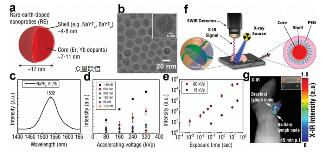
Figure 2. Synthesis and characterization of rare-earth-doped nanoprobes (REs) for X-ray infrared imaging.
a) Schematic of REs showing a lanthanide-doped core surrounded by an undoped shell (NaYF4:Yb/Er@NaYF4);
b) TEM image of REs showing their spherical morphology;
c) X-ray infrared spectrum of REs showing a distinct near-infrared emission peak near 1530 nm after X-ray irradiation;
d) The X-ray infrared detection sensitivity of REs as a function of X-ray accelerating voltage, measured at a constant current of 12.5 mA;
e) Linear distribution map of a simulated phantom cross-section. The exposure sensitivity of the X-ray infrared imaging system was evaluated by exciting REs with 80 kVp and 15 kVp X-rays;
f) Schematic of X-ray infrared imaging of local lymph nodes in mice after footpad injection of PEGylated REs;
g) X-ray infrared imaging of local lymph nodes using PEGylated REs.
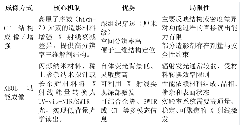
In the imaging dimension, XEOL and CT are not substitutes but complementary: CT is better at providing high-resolution anatomical structural information, while XEOL is better at converting X-ray excitation into functional signals that can be captured by optical systems, for observing luminescent material distribution, energy transfer, or local biological processes. Therefore, Table 1 below is only used to compare the technical positioning of CT structural imaging and XEOL functional imaging in imaging scenarios, and does not represent the full application boundary of XEOL.
Table 1. Comparison of the technical positioning of CT structural imaging and XEOL functional imaging in imaging scenarios
The relationship between capillary lenses and XEOL is not limited to the end of the imaging system. Whether it is spectral acquisition, doping, crystal phase screening, and micro-region luminescence distribution studies on the material side, or deep excitation, low-background readout, and XLCT scanning platforms on the imaging side, all jointly depend on a fundamental condition: converting the divergent light emitted by ordinary laboratory X-ray sources into a controllable excitation beam with higher flux, smaller spot size, and greater stability. Multicapillary X-ray lenses play a role precisely at this stage.
(1) A 1200-fold Flux Leap: Breaking the Brightness Limit of Laboratory Light Sources
XEOL experiments often face a practical problem: the X-ray-to-light conversion efficiency of nanoscintillators is limited, resulting in weak luminescence signals; moreover, ordinary laboratory X-ray tubes output divergent beams, and if only pinhole collimation is used to obtain a small spot, a large number of photons will be blocked, leading to longer integration times and lower signal-to-noise ratios. The value of XOS multicapillary lenses lies in collecting and refocusing divergent X-rays, significantly increasing the photon density per unit area while maintaining spot sizes on the order of micrometers to hundreds of micrometers.
In the research by Professor Changqing Li's team at the University of California, Merced [2]the researchers constructed a focused-beam XLCT system based on the XOS flex-beam. The system used a multicapillary lens to focus the output of a molybdenum-target X-ray tube into an X-ray beam of approximately 100 μm, and compared it with a conventional 1 mm pinhole-collimated beam. The results showed that the luminescence intensity excited by the 0.1 mm focused beam was significantly higher; after normalizing for exposure time, current, and beam spot area, the researchers estimated that the X-ray photon flux density of the focused beam was more than 1200 times that of the collimated beam. This type of flux gain directly improves the acquisition efficiency of weak XEOL signals and makes laboratory-source-based XLCT scanning times more acceptable rather than "very long."
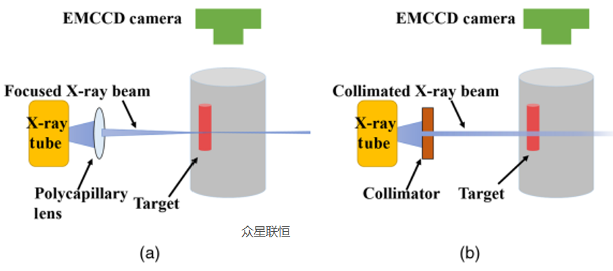
Figure 3. Comparison experiment of luminescence intensity between focused X-ray beam and pinhole-collimated X-ray beam.
a) Approximately 0.1 mm focused beam formed by an XOS multicapillary lens; b) Approximately 1 mm pinhole-collimated beam. The researchers estimated the difference in X-ray photon flux density between the two excitation methods by normalizing the luminescence intensity.
Therefore, the significance of 1200-fold is not just "brighter signals," but more importantly, it transforms laboratory X-ray sources into high-flux excitation sources that can be used for spectral acquisition of weakly luminescent materials and XLCT scanning.
(2) Focused Beam Scanning: Obtaining Micro-Region Luminescence Distribution to Support Material Screening and Imaging Platforms
For nanoscintillator materials, researchers are concerned not only with "what light is emitted and how strong it is," but also with "where the luminescence comes from and whether the distribution is uniform." Overall spectra can evaluate the luminescence intensity and spectral line positions of material batches, but to compare local luminescence distributions under different concentrations, treatment conditions, or tissue coverage, positionable X-ray excitation is required.
In related work by teams at Clemson University and the University of California, Merced [3][4]multicapillary focused beams were used in two types of scenarios: one was XEOL spectroscopy and chemical imaging of NaGdF4:Eu/Tb nanophosphors, where researchers loaded NaGdF4:Eu dispersions of different concentrations into 1 mm glass capillaries and performed scanning with and without tissue coverage to verify that focused X-rays can excite and read luminescence signals at localized positions; the other was a small-animal FXLT platform, which used an XOS fleX-Beam focused source with a focal spot of approximately 50 μm, combined with a translation stage, rotating gantry, and high-sensitivity PMT for optical signal collection.
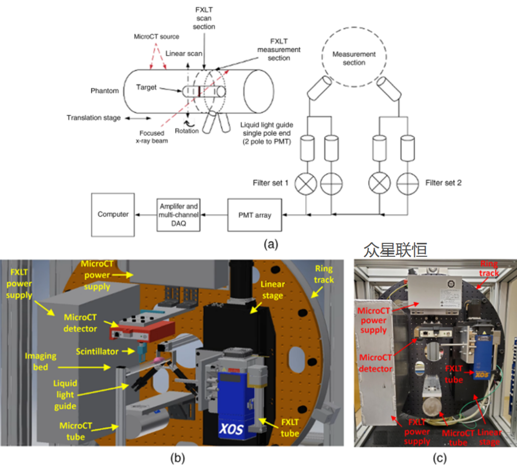
Figure 4. Small-animal FXLT imaging system based on a rotating gantry.
a) FXLT system scanning scheme; b) System CAD model; c) Physical structure of the scanner.
These experiments demonstrate that multicapillary lenses not only "increase brightness," but also define the excitation region: wherever the X-rays are focused, the nanoscintillators there are preferentially excited. For materials R&D, this helps to compare luminescence intensity and spatial uniformity under different synthesis, doping, or heat-treatment conditions; for imaging platforms, localized excitation can provide higher signal-to-noise ratio and clearer spatial constraints for XLCT/FXLT reconstruction.
(3) Focused-Beam XLCT: From Local Excitation to Usable Reconstructed Images
The previous two sections separately illustrated two fundamental advantages brought by multicapillary lenses: one is increasing the X-ray photon flux density, and the other is confining the excitation region to a smaller, more controllable spatial range. However, to make XEOL a truly usable imaging tool, the optical signals obtained from point-by-point scanning must be reconstructed into images with credible positions, sufficient contrast, and even comparable concentrations. This is precisely the step that XLCT/FXLT must cross to move from "can excite, can acquire" to practical application.
Professor Changqing Li's team recently further validated this point [5]The researchers used an XOS molybdenum-target focused X-ray source with a multicapillary optical lens to form a fine beam with a focal spot diameter of approximately 150 μm, and scanned a phantom containing luminescent targets; at the same time, the X-rays transmitted through the sample were also recorded to obtain structural reference images, helping to confirm the true positions of the luminescent targets. To address the issues of signal attenuation with depth in narrow-beam XLCT and the lack of intuitiveness in the reconstruction process, the researchers proposed a weighted sinogram filtered back-projection method that compensates for optical signal attenuation in the projection data, thereby enabling faster and more direct image reconstruction.
In the four-target experiment, the weighted reconstruction results matched the positions in the structural reference images, with a Dice coefficient of 91.5% and a contrast-to-noise ratio of 25.22, both superior to conventional reconstruction; in the lower-concentration target experiment, image contrast was also significantly improved. This demonstrates that the high-flux, positionable focused beam provided by multicapillary lenses not only brightens XEOL signals, but also makes the scanning path and excitation position sufficiently clear, providing the hardware foundation for subsequent fast and usable XLCT image reconstruction.
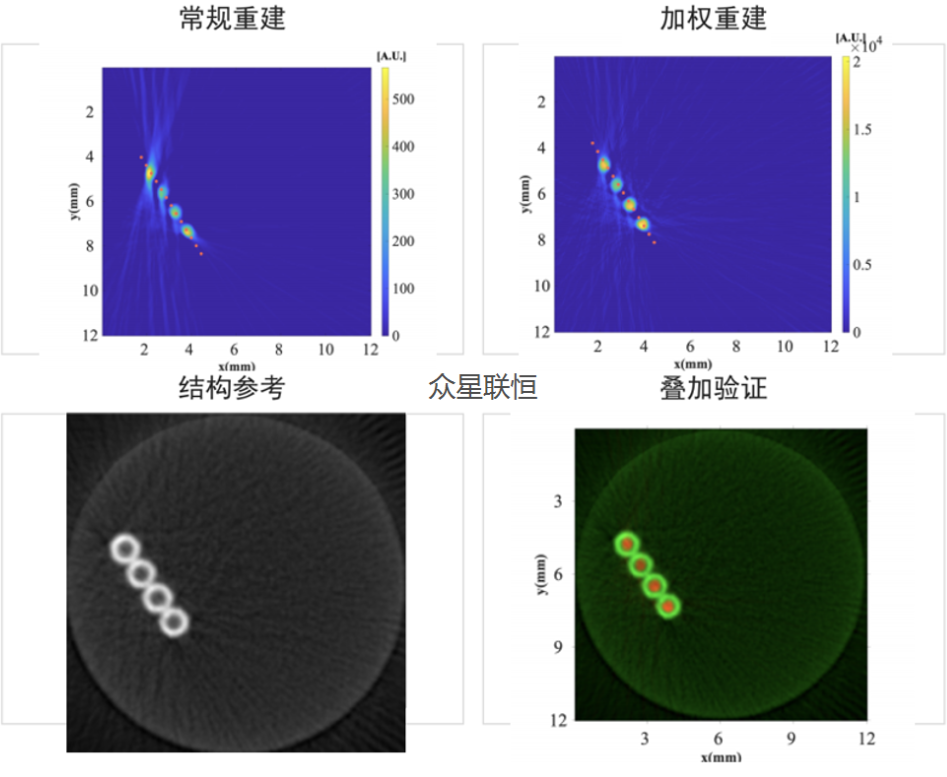
Figure 5. Phantom reconstruction validation of focused-beam XLCT.
The researchers used an XOS focused X-ray source to scan a phantom containing luminescent targets and simultaneously obtained structural reference images. The figure compares conventional reconstruction, weighted reconstruction, structural reference, and overlay validation results, showing that the luminescent targets are reconstructed in the correct regions, with reduced artifacts and improved contrast after weighted processing.
In summary, from the luminescence mechanism of nanoscintillator materials to focused-beam scanning and XLCT/FXLT reconstruction validation, the advancement of XEOL does not depend on a single breakthrough in one link, but rather on a systematic project in which materials, X-ray optics, optical signal acquisition, and image algorithms work together. Materials determine whether sufficiently effective and stable luminescence signals can be generated; detectors and algorithms determine whether weak signals can be read out and restored; and the multicapillary X-ray lens at the excitation end determines whether laboratory light sources can deliver enough X-ray photons to sufficiently small and controllable positions.
This is also the core value of XOS multicapillary lenses in Micro-XEOL scenarios: they increase flux, making spectra and imaging of weakly luminescent materials easier to acquire; they also compress the spot size, enabling researchers to obtain micro-region luminescence distributions and spatial resolution information; and when further combined with scanning platforms and reconstruction algorithms, they provide a practical excitation light source foundation for laboratory-grade XLCT/FXLT systems. For research teams wishing to conduct XEOL material screening, micro-region spectroscopy, or small-animal functional imaging in ordinary laboratories, such optical components are often not merely "icing on the cake," but rather one of the keys to whether the system can work stably.
Of course, XEOL remains a continuously developing technical route: material luminescence efficiency, radiation dose control, system integration complexity, and reconstruction algorithms all still have room for optimization. But these references also point to a clear direction—making the fullest possible use of every X-ray photon and gradually bringing experimental capabilities that previously relied more on large-scale facilities to more laboratory desktops. Top Unistar also hopes to work together with partners such as XOS to provide support for domestic users in building easier-to-use and more stable laboratory-grade X-ray excited luminescence platforms.
The ultimate realization of the value of any cutting-edge technology cannot be separated from deep engagement with localized needs. This is precisely the key role thatTop Unistarplays.
As the long-term strategic partner and general distributor ofAmericanXOScompaniesin the Chinese and Southeast Asian markets,Top Unistaris far more than a channel. We are the "translators," "disseminators," and "practitioners" of technological value. We have a deep understanding of the working principles of capillary X-ray lenses, performance simulation, and the considerations for coupling with X-ray sources, as well as the subtle differences in requirements for capillary X-ray lenses across the wide range of applications involved, such as two-dimensional diffraction XRD, parallel-beam XRD, XRF, full-field microscopy, extreme ultraviolet spectroscopy, and neutron collection, enabling us to quickly recommend suitable specifications to accelerate your research and development process. At the same time, we do not merely introduce equipment; we also place great emphasis on in-depth industry insight, engineering integration support, and continuous localized rapid service. Therefore, we have established a professional technical and market team, as well as anX-ray spare parts inventory(enabling rapid delivery) and ananalytical laboratory(enabling performance demonstration, coupling debugging, and testing), with the ultimate goal of better serving users in various fields.
[1] Fan, Wenpei, et al. "Breaking the depth dependence by nanotechnology‐enhanced X‐ray‐excited deep cancer theranostics." Advanced Materials 31.12 (2019): 1806381.
[2] Zhang, Wei, et al. "X-ray luminescence computed tomography using a focused x-ray beam." Journal of biomedical optics 22.11 (2017): 116004-116004.
[3] Lun, Michael C., et al. "Focused x-ray luminescence imaging system for small animals based on a rotary gantry." Journal of Biomedical Optics 26.3 (2021): 036004-036004.
[4] Ranasinghe, Meenakshi. "Development of Plasmonic and X-Ray Luminescence Nanoparticles for Bioimaging and Sensing Applications." (2022).
[5] Zhang, Yibing, et al. "Weighted Sinogram Filtered Back Projection for Focused Beam Based X-ray Luminescence Computed Tomography." IEEE Access (2026).
Content: Jay·Qiu
Review: Kevin
Layout: Sylvia
Disclaimer:
The content of this article (including images) is partially sourced from the internet. The copyright and views of the quoted portions belong to the original authors. Beijing Top Unistar Technology Co., Ltd. publishes and reposts this content for the purpose of conveying more industry information and network sharing. If you believe this article contains infringing content, please contact us, and we will address it promptly. If you have any questions, please feel free to contact us at any time.
Reprints are welcome; please indicate the source.
FROM READING TO APPLICATION
Put this knowledge to work
Need to assess product suitability, discuss experimental conditions or find technical support? Explore our products and services, or discuss this resource with us.