

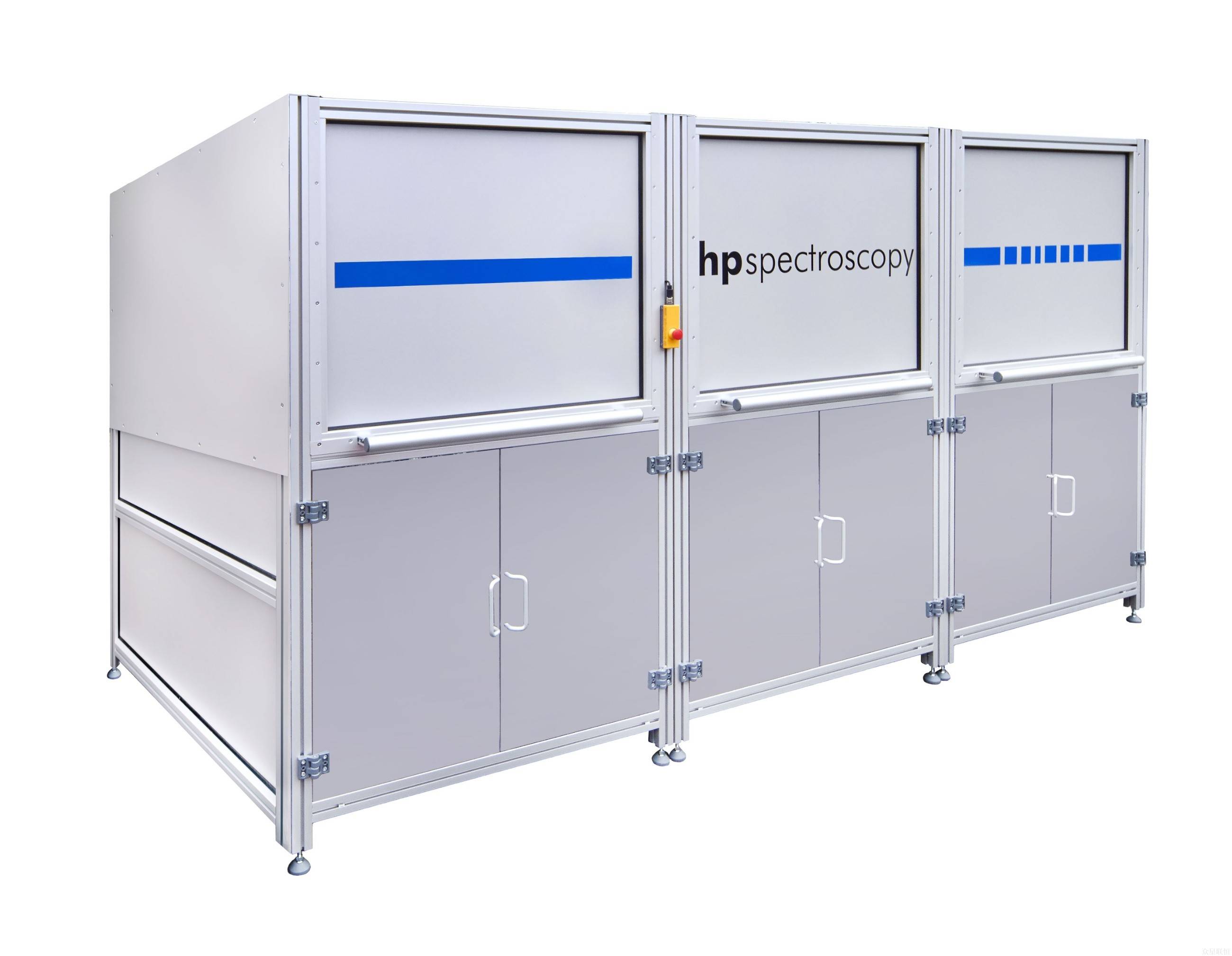

Direct-readout hard X-ray benchtop X-ray absorption fine structure spectrometer 5-12 keV / benchtop XAFS
hiXAS provides a complete solution for laboratory-based extended X-ray absorption fine structure (EXAFS) and X-ray absorption near-edge structure (XANES) spectroscopy. The X-ray tube light source and spectrometer cover an energy range of 5-12 keV.
Model: hiXAS
Brand: HP Spectroscopy

Introduction
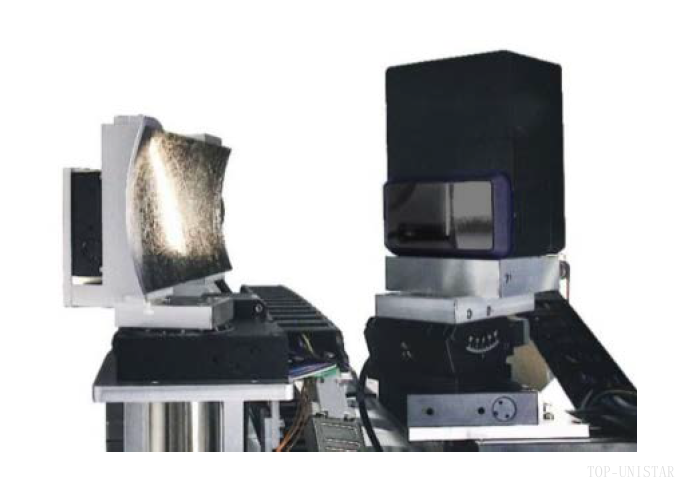
HiXAS provides a complete solution based on laboratory extended X-ray absorption fine structure (EXAFS) and X-ray absorption near-edge structure (XANES) spectroscopy. Within a compact footprint, it integrates an X-ray tube light source, a high-resolution spectrometer, a photon-counting pixelated X-ray detector, and control software for instrument control and data analysis.
Because the spectral quality of hiXAS is comparable to results from synchrotron radiation measurements, the lengthy process of applying for and waiting for synchrotron radiation beamtime is no longer necessary.
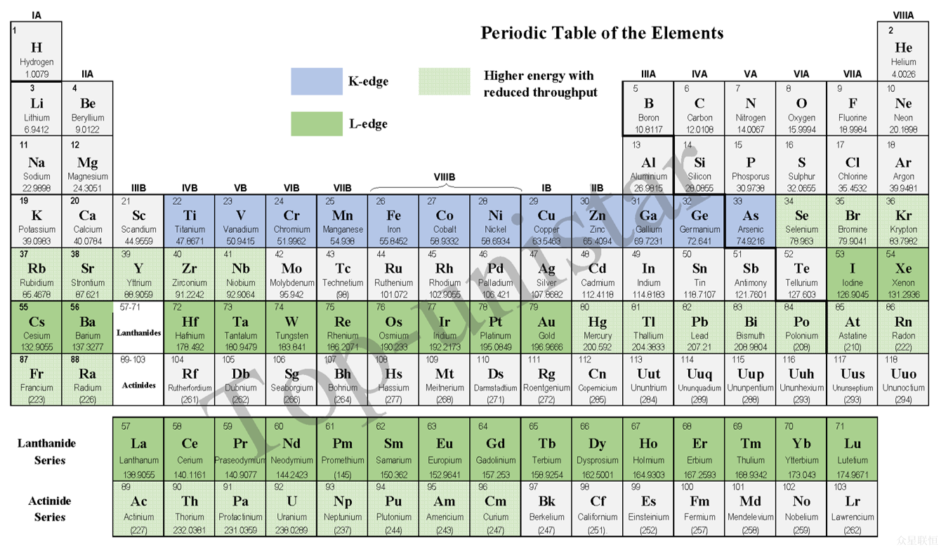
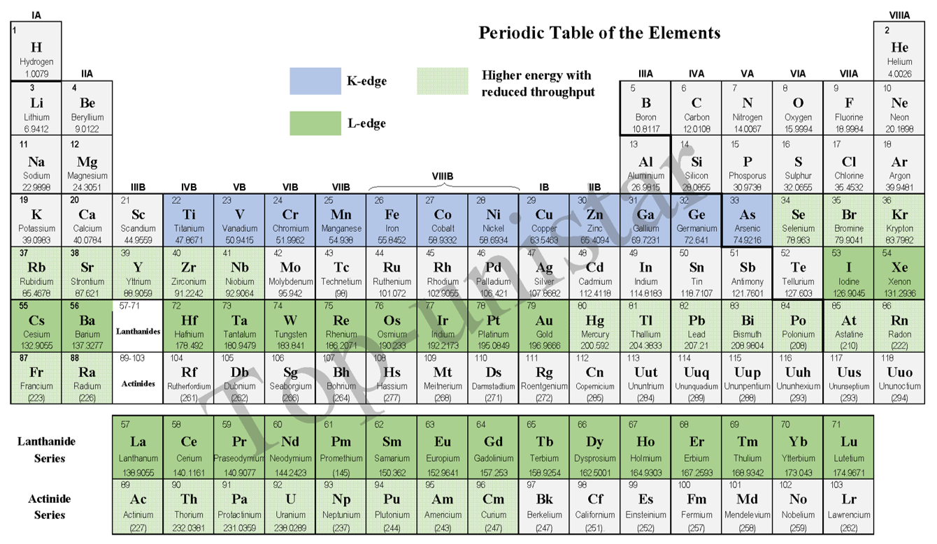
The X-ray tube light source and spectrometer cover an energy range of 5-12 keV, thereby including the K absorption edges of 3d transition metals. The specially optimized HAPG Von Hamos spectrometer geometry enables spectra with exceptionally high signal-to-noise ratios. As a result, sample concentrations as low as a few weight percent can be analyzed. At the same time, the spectrometer maintains high efficiency and a constant high resolution (E/ΔE = 4000) across the covered absorption edge range. We can also provide customized hiXAS systems tailored to your various application needs.
Typical applications
1. Materials science
- Catalyst research
- EXAFS: Analyzing the local structure of catalyst active sites (e.g., coordination number, bond length, and disorder of metal nanoparticles) to reveal the relationship between active centers and reaction mechanisms.
- XANES: Determining the oxidation state (e.g., metal valence) and coordination environment (e.g., octahedral or tetrahedral symmetry) of catalysts, and studying dynamic changes under reaction conditions.
- Application example: Structural evolution of noble metal catalysts (e.g., Pt, Pd) in fuel cells.
- Nanomaterial characterization
- Using EXAFS to study the size, surface atomic coordination, and interfacial structure of nanoparticles; using XANES to analyze the electronic states and chemical bonding characteristics of quantum dots or clusters.
- Amorphous/disordered materials
EXAFS can resolve the short-range ordered structure of amorphous materials (such as glass and alloys), compensating for the dependence of X-ray diffraction (XRD) on long-range order.
2. Environmental and Earth Sciences
- Environmental Remediation
3. Energy Materials
- Battery Materials
- Photovoltaics and Photocatalysis
4. Coordination Chemistry

Specifications
| Core components | X-ray tube light source, von Hamos HAPG spectrometer, photon-counting pixelated X-ray detector | |
| Energy range | 5-12keV | |
| Sample concentration | Down to a few weight percent | |
| Sample mounting | Multiple sample carousel | |
| Footprint | 2.0m x 1.0m (adjustable according to configuration) | |
| Software suite | Integrated system control, various spectral calibration and analysis functions | |
| EXAFS mode | XANES mode | |
| Spectral resolution | 1800* | 4000* |
| (*constant over the entire energy range) | ||
| Energy bandwidth | 1000eV | 300eV |
| Acquisition time | 3 min** | 8 min** |
| (**normalized analyte concentration) | ||
Literature
8 references found
ABSTRACT Conventional non‐noble hydrogen evolution reaction catalysts are plagued by excessive hydrogen adsorption and instability. To overcome this, we pioneer a high‐entropy single‐atom (HESA) catalyst via precise anchoring of multiple transition metals on carbon supports. The HESA catalyst achieves record‐breaking alkaline HER performance of 44 mV@10 mA cm −2 (matching commercial Pt/C catalyst) and 300 h stability, resolving the persistent activity‐stability trade‐off. Systematical characterizations reveal that low‐coordination metal sites induce significant metal‐support charge redistribution. Electronegative supports withdraw electrons from metal centers, while π‐back‐donation downshifts metal d‐band centers, optimizing the Gibbs free energy of intermediate H atoms (ΔG H* ). Atomic‐scale imaging further confirms uniform charge distribution and Ångström‐level electric field response. Crucially, adjacent multi‐metal sites synergistically lower energy barriers for both water dissociation and hydrogen desorption through a synergistic electronic buffering effect. This work establishes entropy‐driven microenvironment engineering as a paradigm for cooperative optimization of active sites and electronic structures, opening avenues for durable non‐precious catalysts.
ABSTRACT In this work, we demonstrate the integration of a flat liquid jet sample delivery system into a compact soft x‐ray absorption spectrometer using a table‐top laser‐induced plasma source. A high‐speed flat liquid sheet is formed by the collision of two cylindrical jets. This micrometer‐thin lamella can ideally be utilized for transmission‐mode soft x‐ray absorption spectroscopy using krypton plasma emission. Detailed analysis of the jet's thickness profile is achieved applying Lambert–Beer's law. Measurements on water, focusing on the oxygen K‐edge, reveal a lamella thickness profile ranging from 500 nm to 1 μm over a length of 3.8 mm. Additionally, we have investigated aqueous solutions of iron salts, capturing near edge x‐ray absorption fine structure spectra over a broad spectral range from the nitrogen K‐edge to the iron L‐edge. Focused analysis on iron species in aqueous solutions enabled us to distinguish quantitatively between the oxidation states of Fe 2+ and Fe 3+ at the iron L‐edge. Our results are compared with measurements obtained under similar conditions at a synchrotron.
Abstract Despite two‐dimensional (2D) conductive metal–organic frameworks (cMOFs) being attractive due to their intrinsic electrical conductivity and redox activity for energy applications, alleviating the constrained mass transfer within long‐range micropore channels remains a significant challenge. Herein, we present a tandem assembly and etching chemistry, to incorporate perpendicularly aligned mesopores into the micropores of cMOFs, via a bi‐functional modulator. Synchrotron spectral and morphological analyses demonstrate that the elaborate ammonia modulator first coordinates with Zn 2+ forming defects during the initial self‐assembly of cMOF oligomers, which then initiates mesoporous cMOFs via in situ etching. In situ spectroscopy and theoretical simulations further reveal that such a unique perpendicular mesoporous structure shorts the micropore channels by two orders of magnitude and relaxes the inherent ion stacking within micropores, leading to five times faster Na + transport and a remarkable rate capability at 250 C and sodium storage lifespan over 50,000 cycles. Our protocol opens up a new avenue for introducing mesopores into microporous cMOFs for advanced energy applications and beyond.
Measurement results and application examples
 | |
 hiXAS enables high-quality data acquisition with an energy resolution comparable to that of synchrotron radiation. |  XANES spectra at the Zn K-edge of single-atom catalysts. High-quality XANES spectra were obtained even at analyte concentrations as low as 1 wt%. hiXAS can measure diluted samples, enhancing the ability to identify oxidation states. |
 hiXAS exhibits high sensitivity to elements and enables analysis of the electronic structure and valence states of elements through the energy position of the absorption edge. |  hiXAS can measure low-concentration samples; the above shows XANES spectra of a Co sample. |
Application examples
  X-ray absorption measurement of a Cu foil sample; acquisition time: 3 minutes with sample, 1.5 minutes without sample. C. Schlesiger et al, Recent progress in the performance of HAPG based laboratory EXAFS and XANES spectrometers, J. Anal. At. Spectrom. 35, 2298 (2020) | |
 (a-b) Comparison of Sm1.5Sr0.5NiO4 material Ni K-edge XANES spectra measured with a laboratory benchtop XAFS spectrometer before and after the DRM reaction; (c-d) Sm1.5Sr0.5NiO4 material Sm L3 -edge and Sr K-edge XANES spectra measured with a laboratory benchtop XAFS spectrometer before and after the DRM reaction. | |
  (b) Co K-edge absorption spectra of Co, Co3O4, and macro-TpBpy-Co measured with the laboratory benchtop XAFS spectrometer hiXAS; (c-d) corresponding R-space spectra obtained via Fourier transform. |