

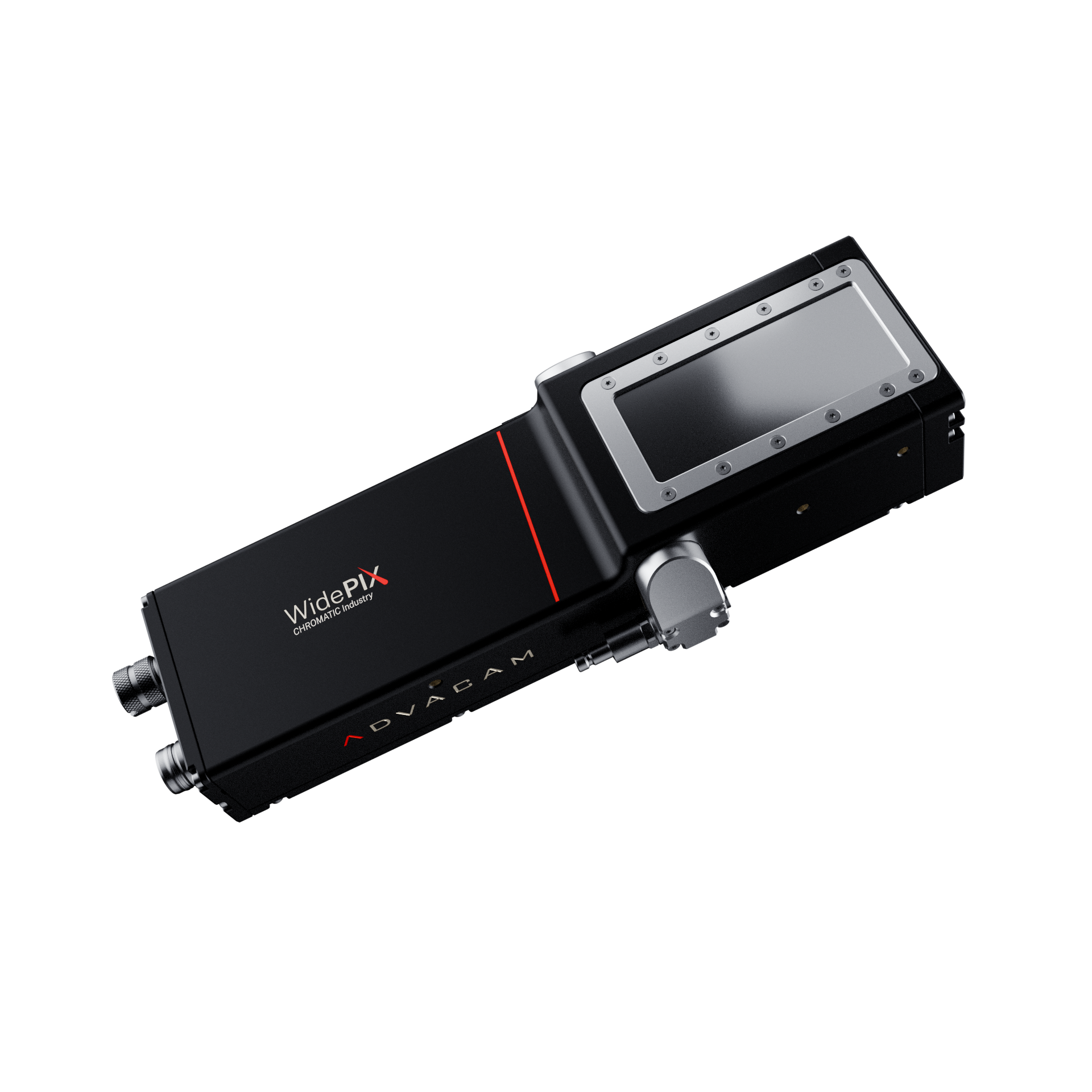
WidePIX CHROMATIC Industry Photon-Counting X-ray Detector
Formerly known as: WidePIX MPX3 PoE. This is a durable, compact, multi-chip scanning and imaging X-ray detector optimized for industrial applications, providing "color radiography" with spectral material sensitivity. It is suitable for CT scanners and supports hardware-based time-delay integration (TDI) mode. Its core uses a Medipix3 hybrid detector with silicon or cadmium telluride sensors. The device achieves simplified connectivity via a single Power over Ethernet (PoE) cable.
Model: WidePIX CHROMATIC Industry
Brand: Advacam

Introduction
The WidePIX CHROMATIC Industry is a multi-chip scanning and imaging X-ray detector. Designed not only for advanced imaging, it is a rugged solution engineered for reliable industrial deployment in demanding environments. It delivers spectrally sensitive, material-sensitive "color radiography," fully compatible with CT scanners, and supports hardware-based time-delay integration mode (single-row configuration only). At its core, it integrates Medipix3 hybrid detector technology with silicon or cadmium telluride sensors.
The detector consists of multiple Medipix3 electronic chip units, each corresponding to a 256 × 256 pixel matrix and perfectly bonded to a silicon or cadmium telluride sensor.
Thanks to its patented near-seamless tiling technology, the entire sensitive area offers full sensitivity, ensuring uniform image acquisition with no significant dead zones (at most only 4 pixels). Each pixel is equipped with two integrated 12-bit digital counters and two energy discrimination thresholds, enabling precise particle counting. The counters can also be merged into a single 24-bit unit to provide a wider dynamic range. This particle-counting method eliminates additional electronic noise, producing high-contrast X-ray images that clearly reveal fine structures such as plastics or soft tissues.
The industrial optimization of WidePIX sets it apart:
Ruggedness — Reinforced structure and optimized electronic components ensure stable operation in continuous industrial workflows.
Universal integration — The compact form factor makes it smaller and easier to handle, and it can be readily installed into confined machine spaces.
Simplified connectivity — A single Ethernet cable carries both data and power, delivering a true plug-and-play experience.
Adaptation to demanding environments — Optimized design with enhanced vibration resistance ensures long-term stable performance under demanding conditions.
Upgraded water-cooling interface — Redesigned for industrial application convenience, accessible from the side of the detector for flexible installation and maintenance.
Spectroscopic imaging capability remains at the core of this system: The dual-threshold energy discrimination technology of Medipix3 enables X-ray spectroscopic imaging, allowing differentiation of materials in a sample based on their attenuation characteristics. The system supports energy-resolved measurements starting from 5 keV (silicon sensor) or 8 keV (cadmium telluride sensor).
Furthermore, the charge summing mode in the pixel electronics corrects inter-pixel charge sharing, significantly improving spectral accuracy and per-pixel spectral quality.
Whether in CT scanners or in continuous scanning applications using TDI mode, WidePIX provides reliable, high-resolution imaging solutions with the robustness and flexibility required by modern industry.
General Parameters
Literature
3 references found
The high-energy X-ray beamline BL15XU is designed to leverage the photon source characteristics of SPring-8 to advance materials science and high-pressure research. The beamline extracts a 100 keV pink beam from undulator radiation via a double multilayer monochromator and delivers a high flux of 6.0 × 10 13 photons/s at the sample position. Experimental hutch 1 is dedicated to materials engineering, employing non-destructive methods based on scanning 3D X-ray diffraction and computed tomography. Experimental hutch 2 focuses on high-pressure science and is equipped with a rotating slit system that enables switching between radiography and diffraction modes at speeds up to 144 Hz, with a minimum exposure time of approximately 2.7 ms. Installed high-pressure equipment includes: a 1500-ton MADONNA multi-anvil press capable of generating pressures up to 120 GPa; a mobile 200-ton Hyaku-shiki press for in-situ deformation experiments up to 20 GPa; and a dedicated Paris-Edinburgh press setup for pair distribution function analysis of liquids and amorphous materials, with a wide momentum transfer range of up to 27.8 Å −1 . These diverse capabilities make BL15XU a versatile platform for in-situ investigations in materials science and high-pressure research, fully prepared for the future high-brilliance operation of SPring-8-II.
In energy-resolved X-ray projection imaging, quantitative effective atomic number ($$Z_{eff}$$) inversion is affected by spectral distortion caused by beam hardening and detector response. This study proposes a joint beam-hardening and detector-response correction framework for thickness-decoupled $$Z_{eff}$$ inversion. By combining the polychromatic X-ray source spectrum, material-dependent attenuation, and the detector response matrix of an energy-resolved photon-counting detector, a folded spectral forward model was established. Based on this model, a response-corrected spectral database was constructed using Monte Carlo simulation. A spectral mass-attenuation linearization method was then employed to reduce the nonlinear attenuation behavior induced by beam hardening, followed by $$Z_{eff}$$ inversion via reliability-weighted least-squares spectral matching. Experimental validation was conducted using standard low-to-moderate $$Z_{eff}$$ materials with theoretical $$Z_{eff}$$ values ranging from 6.5 to 13.0 under four mass-thickness conditions ($$\rho t$$ = 3.0–9.0 g/cm²). The results demonstrate improved thickness stability and quantitative consistency within the calibrated material and thickness ranges. The method was further applied to carbon-fiber-reinforced polymer specimens containing aluminum foil and fiber inclusions. The resulting $$Z_{eff}$$ maps provide material-dependent contrast beyond conventional grayscale attenuation, indicating the potential of the proposed framework for qualitative or semi-quantitative material discrimination in non-destructive testing of composite materials.
Typical Applications
CT imaging
X-ray spectroscopy (XAENS, XES, etc.)
X-ray defect inspection of light materials and soft tissue
Non-destructive testing of large samples
Mineral and geological sample inspection
Industrial radiography and non-destructive testing of high-density components
High-energy X-ray and gamma-ray imaging
Inspection of lightweight mechanical parts and electronic components
Company Introduction
Advacam S.R.O. originated from the Institute of Experimental and Applied Physics of the Czech Technical University, and is dedicated to providing silicon sensor fabrication, microelectronics packaging, radiation imaging cameras, and X-ray imaging solutions across multidisciplinary business areas.
The core technical feature of Advacam is that its X-ray detectors (based on the Timepix chip) have no gaps, resulting in outstanding performance in non-destructive testing, biomedicine, geology and mining, art, and neutron imaging. Advacam maintains strong project collaborations with NASA (National Aeronautics and Space Administration) and ESA (European Space Agency), and its products and solutions are also applied in the aerospace field.
AdVisiones Technologies
It is an imaging technology group that began in science and thrives on ecosystem. Starting from the CERN laboratory, our mission is to make the invisible visible and the impossible possible.
AdVisiones Technologies a.s.
Imaging Innovation Hub.
ADVACAM s.r.o.
Imaging Engine - Focused on the R&D and manufacturing of photon-counting detectors, the source of technology.
Radalytica a.s.
All-in-One Scanning - The world's exclusive mobile robot CT solution.
AdvaScope s.r.o.
Accelerated particle detection - a developer of cutting-edge electron microscope detectors.
InsightART s.r.o.
Deciphering the "genes" of artworks - a cross-disciplinary pioneer in technology and art conservation.
re-medical s.r.o.
Innovators in soft tissue imaging and nuclear medicine - exploring new frontiers in precision medicine.