

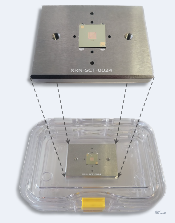
XRN Micro-CT Resolution Test Chart (0.1-50 μm)
X-ray resolution test charts are important tools for measuring the imaging quality of imaging systems. Our company can provide various types of test charts to meet your various needs. Pattern types include line pairs, hole arrays, and Siemens stars.

Introduction
Spatial resolution is an important performance indicator of CT image quality, and its testing is also a key part of CT equipment performance evaluation. The line-pair gauge method, which uses a line-pair test card to directly measure spatial resolution, is widely used in practical industrial CT spatial resolution testing due to its convenience and simplicity.
New-generation micro-CT resolution test card: enhance your X-ray imaging experience! Our cutting-edge test card is meticulously designed and manufactured using modern micro- and nano-fabrication technologies, including top-tier electron beam lithography. The innovative test patterns feature lines and spaces ranging from 50 μm down to 200 nm, as well as Siemens star patterns. Our standard products are designed to exceed expectations. For customers seeking tailored solutions, we also offer fully customizable options.
General Parameters
Literature
2 references found
The utilisation of high-resolution in situ computed tomography (CT) in the (sub-)μm range is typically only viable in synchrotron facilities, as the deployment of a conventional loading stage in laboratory CTs with a cone beam source does not facilitate a corresponding geometric magnification. This publication presents a CT system with a novel in situ concept that allows spatial resolutions down to 0.5 μm, enabling the analysis of weakly absorbing materials capable of applying loads of up to 5 kN in both the compression and tension directions to the sample during the measurement. The necessity for a highly precise mechanical design to ensure successful measurements at magnifications approaching the theoretical limit makes the system’s development particularly demanding. The components employed are presented, along with the requisite considerations and methodologies. It can be demonstrated that the intended specifications with regard to precision and quality are met. The experimental results of a fibre-reinforced polymer demonstrate the system’s ability to detect matrix damage features below a single fibre diameter, thereby highlighting its potential for applications in materials science where traditional laboratory CTs are insufficient and synchrotron access is limited.
Abstract High-resolution x-ray tomography is a common technique for biomedical research using synchrotron sources. With advancements in laboratory x-ray sources, an increasing number of experiments can be performed in the lab. In this paper, the design, implementation, and verification of a laboratory setup for x-ray nano-computed tomography is presented using a nano-focus x-ray source and high geometric magnification not requiring any optical elements. Comparing a scintillator-based detector to a photon counting detector shows a clear benefit of using photon counting detectors for these applications, where the flux of the x-ray source is limited and samples have low contrast. Sample contrast is enhanced using propagation-based phase contrast. The resolution of the system is verified using 2D resolution charts and using Fourier Ring Correlation on reconstructed CT slices. Evaluating noise and contrast highlights the benefits of photon counting detectors and the contrast improvement through phase contrast. The implemented setup is capable of reaching sub-micron resolution and satisfying contrast in biological samples, like paraffin embedded tissue.