

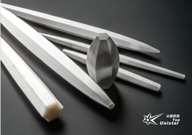
Polycapillary Extreme Ultraviolet and X-ray Lenses
• Integrated with compact low-power supplies, providing flux equivalent to rotating anode sources • Broad spectral bandwidth: 50 eV - 50 KeV • Point-to-point focusing beams • Point-to-parallel beams • Customizable housing designs
Brand: XOS

Introduction
Custom polycapillary X-ray optical solutions are provided based on customer requirements.
Polycapillary collimating lens devices convert highly divergent X-ray beams into low-divergence quasi-parallel beams. These lens devices are primarily used in X-ray diffraction (XRD) and wavelength dispersive spectroscopy (WDS).
Polycapillary focusing lens devices collect X-rays from the source over a large solid angle and focus them into a spot as small as 10 µm. The resulting X-ray flux density is several orders of magnitude higher than that obtained with conventional pinhole collimators. The primary application of these lens devices is micro X-ray fluorescence (XRF) analysis, widely used in thin film and plating analysis, precious metal evaluation, alloy measurement, and circuit board coating monitoring. These lens devices can also be used for detection in applications such as confocal XRF analysis and superconducting energy-dispersive X-ray spectrometers. The use of polycapillary focusing lens devices significantly improves detection sensitivity and allows high performance with low-power X-ray tubes. Micrometer-level spatial resolution enables application in the evaluation of small features in electronics and precious metals. Polycapillary lens devices provide gains of 100x-10,000x, with output focal spots as small as 10 µm.
| Features Order-of-magnitude flux gain from micrometer-sized spots • Integrated with compact low-power supplies, providing flux equivalent to rotating anode sources • Broad spectral bandwidth: 50 eV - 50 KeV • Point-to-point focusing beams • Point-to-parallel beams • Customizable housing designs | 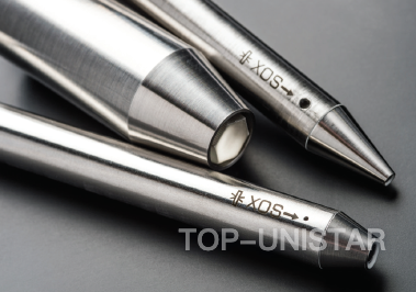 |
Features & Advantages
Specifications
Focusing Optics
| Working distance (mm) | 2 | 4 | 9 | 20 | 50 | 100 | 200 | 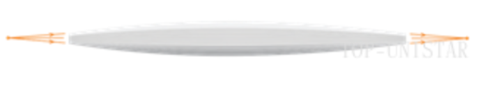 Applications include micro-XRF for elemental mapping, plating thickness and fine feature analysis. |
| Focal spot size* (µm, FWHM, 17.4keV) | 7 | 15 | 25 | 45 | 100 | 180 | 300 | |
| Intensity gain* (vs а plnhole collimator of same size, 100mm fгom the source) | 6000 | 4500 | 3500 | 2000 | 800 | 300 | 120 |
Note: *With а 100μm X-ray source.
| Half-focusing Optics (XRF/XAS) |  Applications include micro XRF, micro XAS, and confocal XRF. Applications include micro XRF, micro XAS, and confocal XRF. | |||||
| Working distance (mm) | 2 | 4 | 9 | 20 | 50 | |
| Focal spot size* (µm, FWHM, 17.4keV) | 7 | 15 | 25 | 45 | 100 | |
| Intensity gain*(vs a pinhole collimator of same size) | 850 | 550 | 400 | 200 | 80 | |
Note: *With an incident beam of 2mm in diameter and a divergent angle of <0.5mrad
| Collimating/Parallel Beam Optics (XRD/WDS/XRF) |  Applications include powder XRD, texture and stress analysis, WDS and confocal XRF. Applications include powder XRD, texture and stress analysis, WDS and confocal XRF. | ||||||||
| Output beam diameter (mm) | 0.5 | 1 | 2 | 3 | 4 | 6 | 10 | 15 | |
| Intensity gain* | 12 | 45 | 130 | 250 | 370 | 470 | 680 | 850 | |
Note: *With а 50μm X-ray source at 8keV, The lFD of the optics is 18mm and the output divergent angle is 0.2 degree.