

Hardware
Product Types Choosing
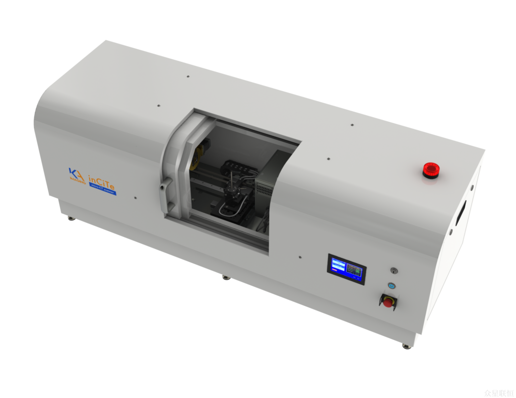
inCiTe™ 3D Scanning Transmission X-ray Microscope / Phase-contrast Micro-CT System
A research-grade phase-contrast X-ray micro-CT system based on KA Imaging’s patented BrillianSe™ high-spatial-resolution amorphous selenium (a-Se) detector technology.
Model: inCiTe™
Brand: KA Imaging

Introduction
inCiTe™3D scanning transmission X-ray microscope — research-grade phase-contrast micro-CT system
Compared with conventional X-ray absorption imaging, inCiTe™ research-grade X-ray phase-contrast imaging provides much greater contrast for light-element samples, making it particularly suitable for soft tissue and specimens composed of light elements. Most of the 5 principal phase-contrast imaging approaches require highly coherent sources and have therefore relied on synchrotron radiation. The large footprint and high construction, operating and maintenance costs of synchrotrons limit their suitability for routine clinical examinations. Research groups worldwide are working to translate phase-contrast methods into clinical practice. Numerical simulations and experiments using conventional X-ray tubes or microfocus sources show that in-line phase-contrast imaging can face long exposure times, limited source coherence and restricted X-ray energy selection.
Features & Advantages
Specifications
Phase-contrast technology — enhanced contrast
Enhanced contrast for low-density materials
 | The inCiTe™ 3D X-ray microscope is a commercial research-grade phase-contrast micro-CT system based on KA Imaging's patented BrillianSe™ high-spatial-resolution amorphous selenium (a-Se) detector technology. The high spatial resolution and detection efficiency of the BrillianSe™ X-ray camera enable rapid phase-contrast imaging and conventional micro-CT in a portable benchtop system. |
Phase-contrast imaging complements conventional absorption imaging. Low-atomic-number materials absorb X-rays weakly and consequently produce low contrast in conventional X-ray images. In these cases, changes in X-ray phase provide a more sensitive imaging signal.
inCiTe™ 3D X-ray micro-CT produces phase contrast through free-space propagation, converting object-induced X-ray phase changes into intensity variations at the detector. This substantially improves contrast when imaging materials that absorb X-rays weakly.
Bell pepper seed test Phase-contrast imaging reveals finer detail in bell pepper seeds


Without phase contrast With phase contrast
Kevlar composite test
A phase-contrast image of a Kevlar composite specimen can be acquired after an exposure of a few seconds.

Individual fibres are visible on the left. The image on the right, magnified 4 times, reveals fibre delamination.
Titanium implant specimen
The images below show an orthopaedic titanium implant within a bone specimen. Phase contrast improves the visibility of the porous bone structure (trabeculae).


Without phase contrast With phase contrast
Biological specimens
inCiTe™ 3D X-ray micro-CT provides high contrast in biological tissue, as illustrated by the mouse knee joint below.

3D rendering
Kidney stone specimen
inCiTe 3D X-ray micro-CT is used to analyse the microstructure of kidney stones. Microstructural features of the stone minerals provide useful data for research into how these stones form.
 |  |  |
| Example slice | 3D rendering | Specimen photograph |
Esomeprazole specimen
X-ray imaging supports pharmaceutical quality control, contaminant detection, packaging-integrity checks, regulatory compliance, and research and development. Non-destructive, detailed inspection of pharmaceutical products can support assessments of product safety, effectiveness and overall quality.
The advanced detector in inCiTe 3D X-ray micro-CT reveals fine structural detail in pharmaceutical specimens.


User-friendly interface
The inCiTe 3D X-ray micro-CT acquisition software provides an intuitive graphical interface for both new and experienced users. The system initialises quickly, allowing a specimen to be scanned less than 20 minutes after the first power-up. inCiTe CT does not require specimen preparation such as contrast agents, staining or sample thinning.